Handling
of Filters and Particle Traps in Correlative Microscopy
JOMESA is
manufacturer of
SEM Filter mounts and SEM particle traps.
|
|
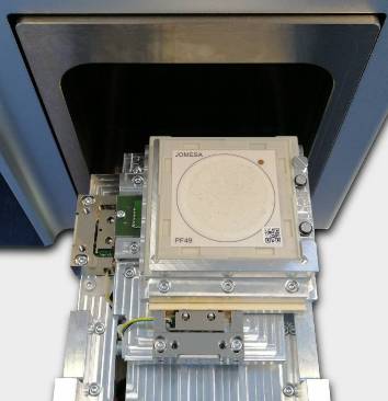
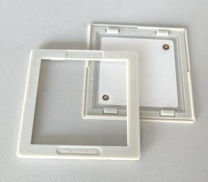
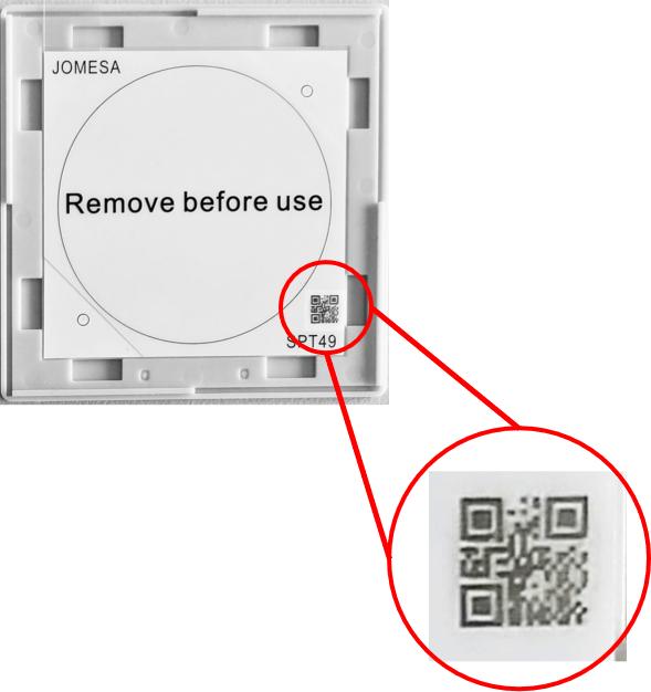
JOMESA SEM Filter
mounts: |
JOMESA SEM Particle
traps: |
|
|
Investigation of filters
without fixation solution is possible only in low vacuum
condition. However the risk of loosing particles by charging
effects is usually not acceptable. Even
in low vacuum fixation is recommended. |
Analysis of particle traps is possible only in low vacuum. |
Specific designed
filter/trap holders allow direct SEM-EDX analysis: |
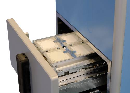
4 Stage PSE |
|
|