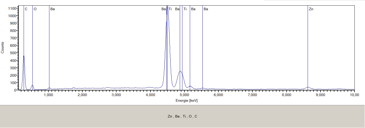
SEM EDX vs. Optical Analysis |
Particles
not (or nearly not) detectable in optical analysis can be found by
SEM analysis and subsequent EDX spectra analysis.
|
|
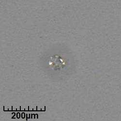
NaCl (Salt) optical image: Particle is not detectable in VDA 19.1 - ISO 16232 frame |
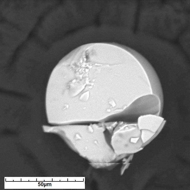
NaCl (Salt) SEM image |
SEM image can look quite different than optical image and can reveal much information |
|
|
|
Optical image |
SEM image |
|
|
|
|
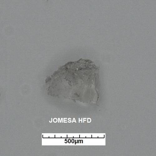
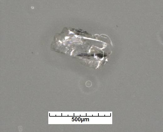
Corundum optical image |
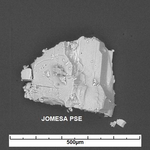
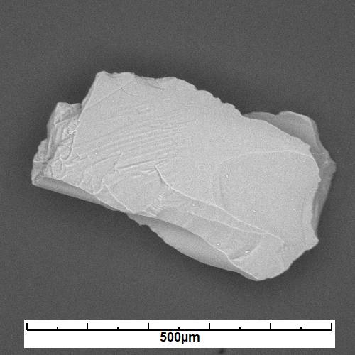
Corundum SEM image |
Critical components (glass, abrasive materials) cannot be detected with optical methods |
|
|
|
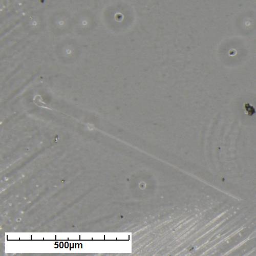
Optical image (glass fiber) |
SEM image (glass fiber) |
|
|
One
cannot compare particle counts of optical and SEM
methods:
These two detection princples
"see" particles different.
Material |
Optical detection |
SEM detection |
Organic (fiber) |
good |
bad |
Graphite |
good |
bad |
Corundum |
bad |
very good |
Glass |
not at all |
very good |
Salts |
bad |
very good |
SEM-EDX versus LIBS (Laser Induced Breakdown Spectroscopy) |
|
|
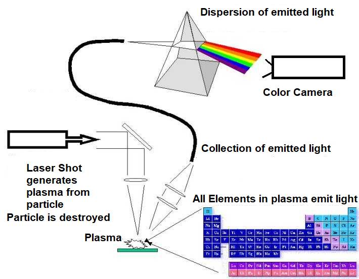
SEM
EDX versus LIBS, Pros and Cons
|
LIBS |
SEM-EDX |
Velocity |
Fast |
Slow (needs vacuum) |
Material depth |
100 µm |
1-2 µm |
Particles |
Only particles visible in optical microscope |
All particles with elements > Boron |
Repeatabilty |
No (particle is destroyed) |
Yes (EDX analysis is non destructive) |
Detection size |
Only particles > 10 µm (laser spot size) |
< 1µm |
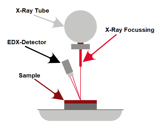
SEM EDX versus XRF (X Ray Flourescence Spectroscopy) |
Originally
XRF is used for bulk material analysis, because X-Rays cannot be
focussed very good.
For
cleanliness/contamination analysis sometimes the technology is
called Hybrid
XRF,
because also image analysis will be used to identify contamination
particles.
First
step: detection of particles. Light elements will generate only faint shadows, heavy elements will generate black spots. |
|
|
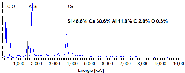
Next step: spectrum analysis of particles |
|
|

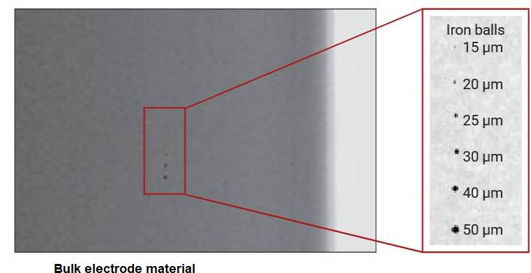
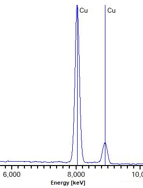
For application where heavy elements (Fe, Cu…) of sizes > 20µm have to be found in large bulk areas, Hybrid XRF is ideal.
Due to limited spacial resolution, smaller particles and particle structures are not detectable.